Hip PAO
The operation of periacetabular osteotomy is designed to move the socket (acetabulum) of the hip joint so that it covers more of the femoral head (ball). The aim is to improve the biomechanics of the hip joint and reduce the high stresses that start to cause damage and arthritis because of the shallow acetabulum. Sometimes it is recommended that an arthroscopy of the hip is undertaken sometime prior to the PAO. This allows the changes within the hip joint to be accurately assessed and and frequently procedures can be performed to the cartilage to optimize the outcome of the PAO.
The operation is usually performed either under a spinal anaesthetic with sedation, or a general anaesthetic with an epidural in place. The surgery itself usually takes about 2 hours. The incision is curved over the outside of the pelvic bone and is typically 10-12cm in length. The operation involves a series of bone cuts around the acetabulum, freeing it from the pelvis and allowing it to be moved to a new position. The new position of the joint is checked with X-rays at the time of surgery and once the optimal position for the hip has been achieved, it is fixed in place with 3 or 4 screws.
After the surgery, patients are mobilized out of bed with the aid of the physiotherapists the day after the operation. Pain is initially controlled with a self administered morphine pump (PCA – patient controlled analgesia). The aim is to get safe on crutches only putting a small amount of weight through the leg. On the third or fourth day following surgery exercises are started in the hydrotherapy pool. Hospital stay is usually 5 to 8 days.
The aim of the surgery is to improve the pain coming from the hip. It is also anticipated that the risk of the development or progression of arthritis will be reduced in the long term, however, this depends a lot on how much arthritis damage has already occurred to the joint before surgery.
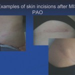
 The original incision and surgical approach to perform a PAO was quite long and extensive. Overtime we have gradually developed a new approach minimizing the dissection and release of soft tissues and in addition making the skin incision only 8-10 cm in length. The skin incision is positioned in such a way that it heals very cosmetically and would not be visible with beachwear.
The advantage of this sort of approach is that the immediate recovery is somewhat quicker and allows more rapid rehabilitation. Usually we allow patients to put about 30Kg of pressure on the operated leg after surgery which makes mobilizing with crutches much easier. After 6 weeks full weight bearing is allowed with gradual progression off 2 crutches onto 1 crutch.
Clearly, a periacetabular osteotomy is not a small procedure. However, with appropriate experience it can be performed through a relatively small incision with a low risk of complications and with a predictable end result.
Because the pelvic bone has a very good blood supply and is surrounded by a lot of blood vessels there is the potential for significant bleeding to occur. During the surgery itself, a cell saver is used which allows blood lost in the wound to be given back to the patient during the operation. It is rather rare for patients to require a blood transfusion.
Because the acetabulum is surrounded by a lot of important nerves and blood vessels there is a small risk of damage to one of these serious structures. This could lead to some weakness in the lower leg. The overall risk of a major complication such as this is in the region of 2%.
Deep vein thrombosis and pulmonary embolus. During the recovery you will be treated with blood thinning injections and thromboembolic deterrant stockings to reduce this risk (2%).
These may occur about 1% of the time and may require antibiotics or further surgery to deal with them.
Failure of the osteotomy to unite. Because the pelvic bone has such a good blood supply and muscle cover, it would be very unusual for the osteotomy not to unite. If this were to happen then further surgery may be needed to stimulate the bone to heal.
Because of the location of the incision for this operation and because of the anatomy of specific small sensory nerves, there is always some numbness over the upper outer aspect of the thigh. This area gradually gets smaller with time. Rarely the numbness may affect the outer aspect of a major portion of the thigh extending down to the knee but it would be unusual for it to persist to this extent.
In time the joint may become arthritic and this depends on the extent of damage before surgery and how shallow the acetabulum is. Although the aim is to make the hip look as normal as possible, it still won’t be a normal hip and therefore the stresses through it may result in the development of arthritis at some stage.
After discharge from hospital it is very helpful to continue with hydrotherapy for 6 weeks after the operation, usually 2-3 times per week. This allows strength and range of movement to be worked on while weight bearing through the hip is restricted. It is worth investigating prior to surgery whether a hydrotherapy facility is available local to you so that this can be organized in advance.
After 6 weeks patients are reviewed and a repeat X-ray taken to ensure that the bone is healing satisfactorily. At this point half body weight is allowed through the leg using 2 crutches for 1 week and then full weight bearing is allowed graduating onto 1 crutch. The exercise programme in this phase of the recovery concentrates more on strengthening the muscles around the hip in particular the hip abductors (gluteus medius and minimus). These muscles need to be strong to avoid any limping when taking full weight through the leg. Patients usually find they can return to most activities 10-12 weeks after surgery and to impact exercise and sports after 6 months.
In general there are no restrictions with activity level after a PAO, but the kinds of sports undertaken should take into account any arthritic damage that may have taken place prior to the osteotomy.
The rehabilitation after a PAO tends to progress in 6 week periods.
0-6 weeks post-op
The focus during this period is to generally increase the patient’s mobility using crutches and working on active movements of the hip.
Weight bearing is limited to 20kgs during this period
Patients should be encouraged to walk with a normal heel-toe gait checking the amount of pressure through the leg by pressing down on some weighing scales.
Hip movements to encourage in the standing position are flexion, abduction and extension exercises.
There is often a lot of inhibition of the hip flexors early on because of the location of the pubic osteotomy
Hydrotherapy is an excellent way of assisting the recovery of these movements but is not essential
Lying down and sliding the heel up and down is a useful way of encouraging active hip flexion.
After 4 weeks, further exercises are built in to the programme.
Side lying hip abductor raises with the hip and knee extended.
If this is difficult at the start, often beginning with clam abductor exercises is helpful and then progressing onto raising the whole leg. Aim to hold the leg in the abducted position for 10 seconds. This aim is to build up to 25 repetitions twice per day
Passive hip flexion towards the chest.
The knee should be pulled and held toward the chest at the point that it becomes rather sore. Hold at that point for 10 seconds. This should be repeated 20 times per day. Everyday passive hip flexion will gradually increase.
Straight leg raise exercises
These can be started against gravity and the aim is to raise the leg approximately 30 cms and hold for 10 secs. This should be repeated 15 times but not more as the ilio-psoas can become rather irritable.
Prone lying hip extension
It is good also to focus on hip extension to stretch the iliopsoas which can become rather tight and to activate the posterior gluteal musculature.
Hip adductor strengthening
As well as working on the hip abductors it is also important to activate and strengthen the adductor musculature. This is best done sitting with a bolster between the knees and squeezing for 10 secs and repeating.
6-12 weeks
At 6 weeks patients will have a check X-ray to ensure that union of the osteotomy is progressing satisfactorily. At this point weight bearing is increased to 30Kgs. This should continue for 2 weeks. At that point patients can be 50% PWB for a further week using 2 crutches. They can then progress onto using 1 crutch. They should continue using 1 crutch until their Trendelenberg gait disappears.
During this time the focus is on progressive muscle strengthening and stretching exercises. The same muscle groups as above need to be worked on but some resistance can be gradually built in such as using therabands.
At 8 weeks using an upright stationary exercise bike is helpful. This helps improve hip flexion and will improve overall hip mobility. Light resistance and around 50-60 rpm is suitable. The duration can be gradually increased aiming to do 30 minutes. From 10 weeks the resistance and rpm can be increased as comfort allows.
Using a cross-trainer from about 10 weeks post op can also be a helpful tool to aid recovery.
If patients have access to a swimming pool, they should be encouraged to start some swimming at 8-10 weeks and to be exercising the hip in the water as well as on land.
12 weeks onwards
At this point the pelvic osteotomy will have consolidated. The pubic osteotomy can take longer to unite depending on the correction required for the acetabulum; the greater the correction the greater the displacement of the pubic osteotomy and so the longer this takes to unite – sometimes even up to 18 months. This by itself is not an issue, but the callus around the osteotomy and the change in the contour of the pelvic brim can affect the iliopsoas tendon and continue to make this irritable. Patients may continue to experience discomfort in the groin region on SLR activities.
The focus should be to continue to work on stretching and strengthening of the iliopsoas, this maybe better standing as opposed to supine so as not to overstress the tendon during strengthening work.
Hip abductor and adductor strengthening work should continue to build up the stamina of these muscles. Often patients will describe that their limp returns towards the end of the day with early fatiguing of the muscles.
Depending on comfort levels progress with increasing resistance work can continue.
For those wishing to return to sports activities, it is important to build up with a low impact exercise programme using the stationary bike, cross trainer and swimming where possible. At 5 months some impact exercises can be introduced and gentle jogging for short periods. This can be increased as tolerated from 6 months onwards.
The procedure itself usually takes about one and a half to 2 hours.
Usually the hospital stay is somewhere between 3 and 7 days. It all depends on how easy you find it to get moving and start using crutches. We aim to get you out of bed the day after surgery and we are happy for you to leave hospital as soon as you are independently mobile.
Over a number of years we have developed a minimally invasive way of doing the PAO. The incision is 8-10 cm and usually in the bikini line so is very cosmetic.
There is almost always a labral tear in patients who have become symptomatic with hip dysplasia. Once the position of the acetabulum has been addressed the load going through the labrum is changed and this then is no longer a source of pain. There will be some occasions where we feel this may need to be addressed particularly if associated with an abnormal shape to the femoral head. If this is the situation then it may be necessary to undergo a hip arthroscopy as well. If this is anticipated in advance then we will often do this about 8 weeks after the PAO and this then won’t interfere too much with the overall recovery from the PAO
The overall length of time on crutches may be up to 10 -12 weeks. The first 6 weeks will feel rather slow in that only 20Kgs of weight is allowed through the leg. After 6 weeks this increases to 30Kgs and from about 9 weeks from surgery you can start to use 1 crutch rather than 2. It will be necessary to use 1 crutch until the tendency to limp disappears which is very much related to building up the strength of the muscles around the hip.
During the time on crutches it is common for people to notice that the foot can be quite discoloured and swollen; often rather red/purple in colour. This is because when using crutches the foot muscles and calf muscles are not being used properly. The best way to minimise this is to be vigorous with calf and foot exercises and when walking to make sure that you are going from heel to toe even though only putting 20Kgs of pressure through the foot.
It can be tough on the hands being on crutches for a significant period of time. The pressure on the heel of the hand can also lead to local tenderness and because there are some nerves that supply sensation to your fingers in this location, it is not uncommon to feel some numbness and tingling in certain parts of the hand and fingers. Wrapping the handles of the crutches can be very helpful, and I recommend tennis racquet grips which tend not to slip around and can be applied to a thickness that suits you. They are cheap and easily available online or at sports shops.
From the mobility point of view this is not specifically necessary, however, it is hard work moving around on crutches and hands tend to get sore. It can be helpful to rent a wheelchair for a few weeks and then it is easier for friends and family to take you out and about and generally less tiring. Wheelchair rental is easy and you can look online to see what is available in your area.
From about 8 weeks onwards there are exercise activities that can be pursued in the gym. In particular using an upright exercise bike. I usually recommend light resistance and not more than about 50-60 rpm to start with. Other activities such as using a cross trainer or treadmill for walking would usually be from about 12 weeks post op. Continuing with a low impact exercise programme and Pilates-type exercises is worthwhile. Impact exercise such as jogging is usually possible to start from about 5-6 months from surgery.
This depends a lot on the type of work you do. In general I recommend warning your employer that you are likely to need about 3 months off work. If you do a sedentary job and can get to work easily then often 10 weeks is OK. It also depends if you can return to work still using crutches. Working from home is certainly possible and anytime from about 6 weeks would be reasonable. If your job is very physically active or you have a difficult commute on public transport, then you may need about 4 months off work.
If the left leg has been operated on and you have an automatic car, then probably from about 6 weeks. If you have a manual car with a clutch, then this is likely to be from about 8 weeks. If the right hip has been operated on then usually from about 8 weeks.
This usually depends on comfort levels rather than any specific point in the recovery from surgery, but usually will be from around 6 weeks after surgery.
Most of the recovery from surgery will take 4-6 months, but it often takes longer for the hip to be fully recovered. This is because it takes the pelvis quite a long time to re-model after this surgery and while this is going on there can be a background level of achiness which is apparent. This will gradually lessen with time.
The PAO is aimed at improving the pain you get from your hip. By re-positioning the acetabulum the stresses through the joint are made more normal so that the tendency for the joint to be damaged and develop arthritis changes will be decreased. If you already have some arthritis in the hip or the shape of the hip is particularly shallow then the joint will always be somewhat compromised and ultimately may require a hip replacement if the arthritis changes progress. We would hope that this would be some way off in the future and would be significantly delayed compared with not having the PAO
There is no reason why pregnancy should be specifically affected after a PAO. There is no contra-indication to natural childbirth
The screws used to hold the position of the osteotomy can become prominent where the screw heads lie and become rather uncomfortable. If this is the cases then the screws can easily be removed. This is done as a daycase procedure under a general anaesthetic. There is no specific need for crutches afterwards and return to normal activity can occur within a few days. Screws can be removed anytime from about 6-8 months after surgery. The process of screw removal does not cause any weakening of the bone. If the screw heads do not become prominent then there is no specific need to have these removed


